Figure 1
Bronchoscopic finding with the use of flexible bronchoscope. It shows a huge tumour that causes almost complete obstruction of the tracheal lumen.

Figure 2
Bronchoscopic photograph, through a flexible bronchoscope after the use of Nd YAG Laser. 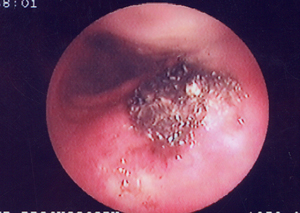
Figures 3a-b
Surgical specimen. Segment of the trachea invaded by a malignant tumour
(at the middle of the specimen). The length of the resected trachea measured about 4.5 cm in length.
A-B
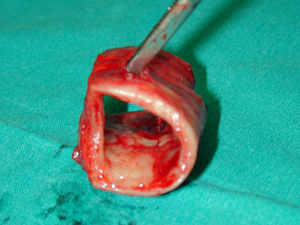
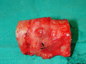
Figure 4
Bronchoscopic photograph through a flexible bronchoscope after the tracheal resection and reconstruction. The suture line is visible and the suture material used was a non-absorbable, monofilament suture. The type of suture is continuous in one layer without any need to reinforce the external suture line. 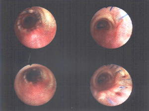
Top