Figure 1
Postero-anterior chest X-ray. There is a shadow in the right upper lobe. Bronchoscopy showed a mass obstructing the orifice of the right upper lobe bronchus.
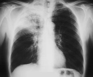
Figure 2a-b
Chest CT scan at different levels. Atelectasis, with inflammatory changes are observed at the right upper lobe because of a mass that infiltrates and obstructs the orifice of the right upper lobe bronchus.
A-B
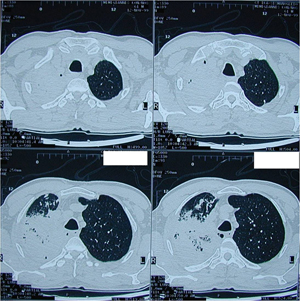
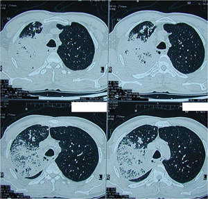
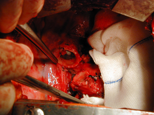
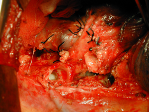
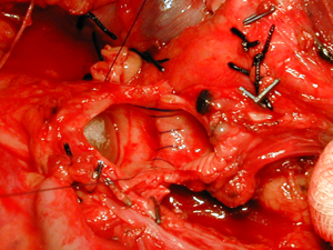
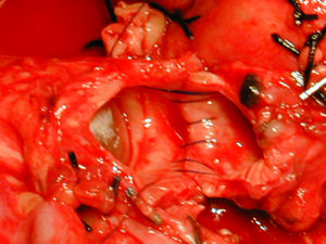
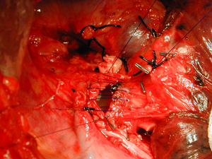
Figure 3a-g
Intra-operative photographs after a right sleeve upper lobectomy. The defect in the tracheo-bronchial tree is shown. Gradual approximation of the two ends of the bronchial tree and an end to end anastomosis was performed.
A-B
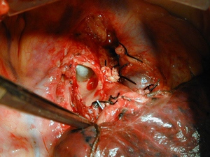
C-D
E-F
G

Figure 4
Postero-anterior chest X-ray after a right upper sleeve lobectomy. Complete expansion of the middle and lower lobe, five years postoperatively.
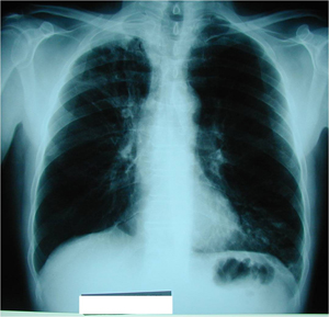
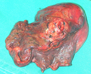
Figure 5
Surgical specimen photograph. A right upper lobe is shown with a tumour at the orifice of its lobar bronchus. The histology examination showed a Non Small Cell Lung Cancer.
Top