Case 1
Figure 1
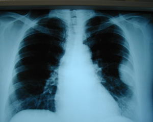
Chest X-ray (postero-anterior), which shows an opacity in the left hemithorax, at the lower lung field.
Case 1
Figure 2
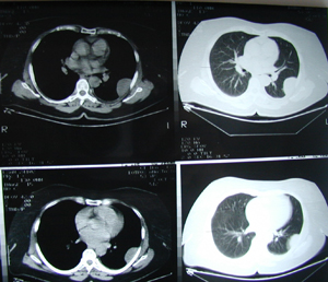
Chest CT scan which shows a tumour mass at the lower lobe, with smooth borders and with no calcification.
Case 1
Figure 3
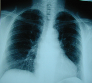
Postoperative chest X-ray after the resection of the lung mass through a mini thoracotomy. The final histology examination showed a rare lung tumour
(sclerosing haemangioma).
Case 1
Figure 4
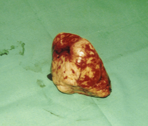
Photograph of the surgical specimen, represents the resected tumour, which was removed by means of a wedge resection.
Case 2
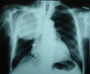
Figure 1
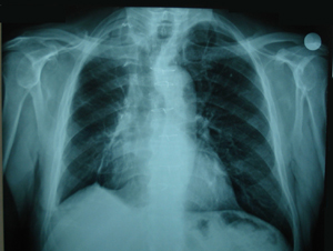
Postero-anterior chest X-ray. There is a huge mass which occupy the right upper and middle lung fields.
Case 2
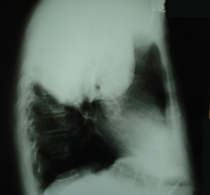
Figure 2
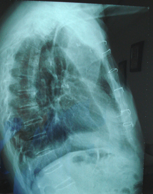
Lateral chest X-ray. There is a huge mass in the upper lung field.
Case 2
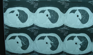
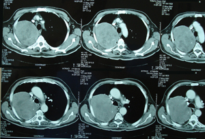
Figure 3a-d
Chest CT scans at different levels. It shows a huge tumour mass with some necrosis, that displaces the mediastinum, to the left side, without any sign of invasion of the large vessels. This mass seems to have smooth borders.
A-B
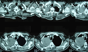
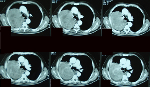
C-D
Case 2
Figure 4.
Postoperative, Postero-anterior chest X-ray. A right upper and middle lobectomy was performed, through a right anterior thoracotomy and median sternotomy. There is a complete expansion of the remaining right lower lobe.
Case 2
Figure 5
Postoperative lateral chest X-ray, after a right thoracotomy and median sternotomy. Normal postoperative image.
Case 2
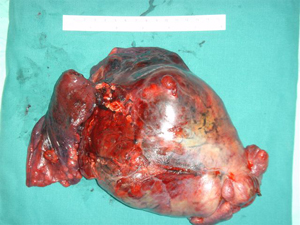
Figure 6.
Surgical specimen photograph, which shows the right upper lobe, the middle
lobe with the huge tumour. The tumour dimensions were 17,5 x 15 x 11,5 cm.
and the histology examination of the specimen revealed a very rare tumour
which was an intrapulmonary solitary fibrous tumour.
Top