Figure 1
Postero-anterior chest X-ray which shows a total pneumothorax of the right hemithorax, and the air displaces the mediastinum to the left side.
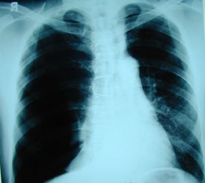
Figure 2
Chest X-ray after the placement of α chest tube. The lung is completely expanded while an extensive subcutaneous emphysema is presented because of functional insufficiency of the chest tube, which is placed in the right position.

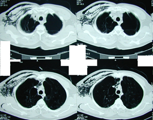
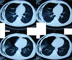
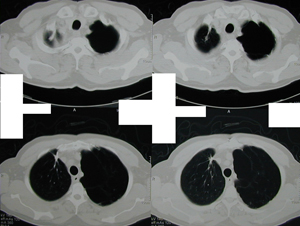
Figure 3a-e
Chest CT scans at different levels showing a large pneumothorax with subcutaneous emphysema. Also there are multiple lung air-cysts of different sizes, at the superior segments of the upper lobes in both lungs.
A-B
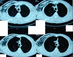
C-D
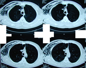
E
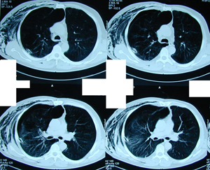
Figure 4
Chest X-ray postoperatively. Complete expansion of the lung after the air cyst resection and lung suture at the site of air-leak.
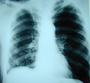
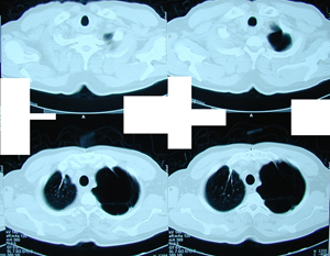
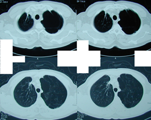
Figure 5a-c
Postoperative chest CT scans at different levels. Normal image of the right lung, while it remains unchanged on the left side where the emphysematic cysts continue to exist.
A-B
C
Top