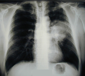
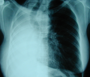
Case 1
Figure 1
Postero-anterior chest x-ray which shows a huge hilar mass in the left lung. The patient was a male heavy smoker.
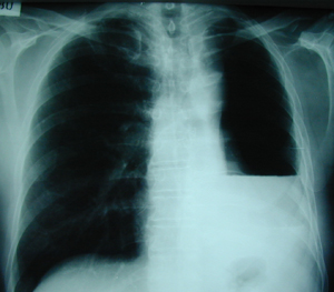
Case 1
Figure 2
Postoperative chest X- ray, one month after the operation ( Pneumonectomy ). There is an accumulation of fluid and air in the left hemithorax (air-fluid level), which is a normal and expected radiological finding. During a period of three weeks to seven months postoperatively ( median 3.9 months) the whole cavity is filled with fluid. The mediastinum is shifted to the operation side, the hemi-diaphragm is elevated and the intercostal spaces are decreased. This procedure results in the limitation of the pneumonectomy space which allows the remaining lung to hyper-inflate.
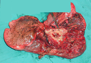
Case 1
Figure 3
Surgical specimen which shows an entire lung with a tumour, located in the upper lobe and extended to the hilum. The pathology examination confirmed the diagnosis of squamous cell carcinoma, which represents the most frequent histologic type .
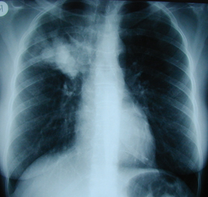
Case 2
Figure 1
Postero-anterior chest X-ray. There is a huge mass located in the right upper lobe.
Case 2
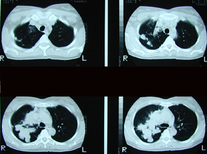
Figure 2
Chest CT scan showing a huge lobulated tumour-mass of the right upper lobe. The mass extends to the lung hilum.
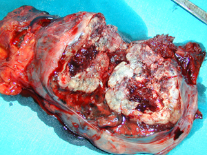
Case 2
Figure 3
Surgical specimen. An entire lung with a large tumour in the upper lobe which seems to invade the hilum.
Case 2
Figure 4
Postoperative chest X–ray after a right pneumonectomy many years after the operation. There is an over expansion of the left lung, while the right hemithorax is filled with fluid which is the expected postoperative image.
Top