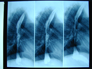
Figure 1
Oesophageal X-ray after barium swallow meal. There is a distention of the oesophageal lumen above a filling defect of the oesophagus.
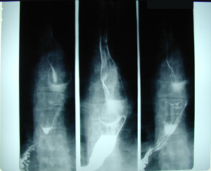
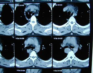
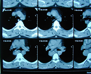
Figure 2a-c
Chest CT Scan. There is a tumour at the posterior mediastinum, probably originating from the oesophageal wall.
A-B
C
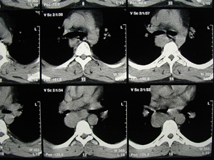
Figure 3
Photograph of the surgical specimen. A tumour of the oesophageal wall has been resected, and it is a horse-shoe shaped tumour. The histology examination revealed a Leiomyoma of the oesophageal wall, which represents a very rare pathologic entity.
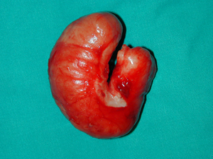
Figure 4
Intra-operative photographs through a limited right postero-lateral thoracotomy, showing the oesophageal tumour being resected.
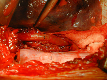
Figure 5
Oesophageal X-ray postoperatively, after a barium swallow meal. Normal oesophageal lumen.
Top