Case 1
Figure 1
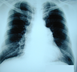
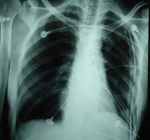
Postero-anterior chest X-ray. It reveals a mediastinal widening which is caused by a huge mediastinal mass.
Case 1
Figure 2
Lateral chest X-ray. It shows a shadow located retrasternally.

Case 1
Figure 3
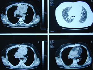
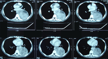
Chest CT scan, that confirms the chest X-ray findings. A huge tumour mass is located anteriorly to the pericardium (anterior mediastinum) and posteriorly to the sternum. The most frequent histologic type at this location is a thymus gland tumour.

Case 1
Figure 4
Chest CT scan of the same patient at a lower level.
Case 1
Figure 5
Postero-anterior chest X-ray after the resection of the tumour through a median sternotomy. The histology examination showed an ectopic thyroid tissue from which this huge tumour has originated.

Case 2
Figure 1
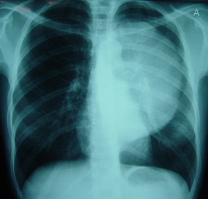
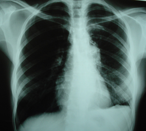
Postero-anterior chest X-ray which shows a huge mass located at the posterior mediastinum.
Case 2
Figure 2
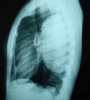
Lateral chest X-ray. There is a huge mass at the posterior-mediastinum.
Case 2
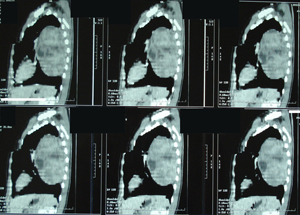
Figure 3a-d
Chest CT scan at different levels, that confirms the findings of the chest
X-ray. There is a huge tumour mass located at the posterior mediastinum, without any sign, suspicious of the adjacent structures, infiltration.
A-B

C-D

Case 2
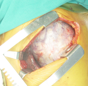
Figure 4a
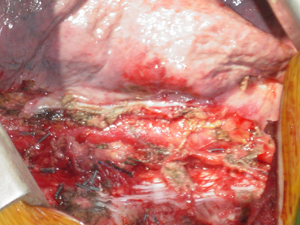
Intra-operative photograph. After the retraction of the ribs, a mass emerges through our incision, originating from the mediastinum.
Case 2
Figure 4b
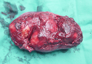
Photograph of the surgical specimen. A mass that measures 17 x 10.8 x 9.5 cm. The histology examination showed a benign neurogenic tumour,
( shwannoma or neurolemoma ).
Case 2
Figure 4c
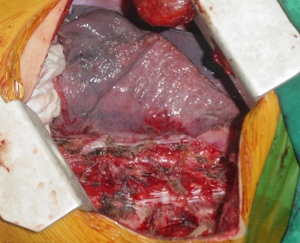
Intra-operative photograph after the resection of the mediastinal tumour. The atelectatic lung is visible.
Case 2
Figure 4d
Intra-operative photograph after the resection of the mediastinal tumour, with the lung being inflated.
Case 2
Figure 5
Postoperative postero-anterior chest X-ray after the resection of the tumour through a left lateral thoracotomy. The two chest tubes are visible and the lung is expanded completely.
Case 2
Figure 6
Postero-anterior chest X-ray, one month postoperatively.
Complete expansion of the lung without any abnormality.
Top