Figure 1
Postero-anterior chest X-ray that reveals the existence of a shadow in the right lung, of a male patient heavy smoker.

Figure 2
Lateral chest X-ray. A huge mass located in the right upper lobe, at the aortic arch level.
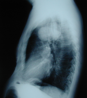
Figure 3
Chest CT scan that confirms the chest X-ray findings. It shows a lobulated tumour-mass located in the right lung.
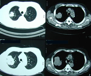
Figure 4
Postero-anterior postoperative chest X-ray, three days after right upper lobectomy. Complete expansion of the remaining lung. Also chest – tube is visible in the right hemithorax.
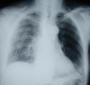
Figure 5
Postero-anterior chest X-ray, many years after the right upper lobectomy. Almost hardly could someone realize that an operation – lobectomy, had been performed.
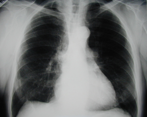
Figure 6
Lateral chest X-ray. Normal image, many years after a right upper lobectomy. The lobectomy was performed as a treatment for non small cell lung cancer.

Figure 7
Photograph of a resected lung lobe with a tumour into the lung parenchyma
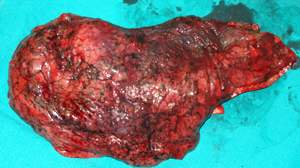
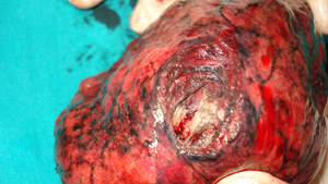
Figure 8
Photograph of a resected lung lobe. An incision has been made above the tumour, which is located deeply into the lung parenchyma.
Top