Case 1
Figure 1
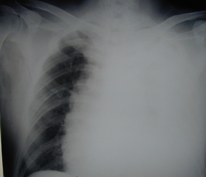
Chest X-ray showing massive collection of fluid in the left hemithorax, which causes displacement of the mediastinum to the right side. The patient presented severe symptoms of dyspnea and discomfort. Also he was on anti-coagulant medication.
Case 1
Figure 2
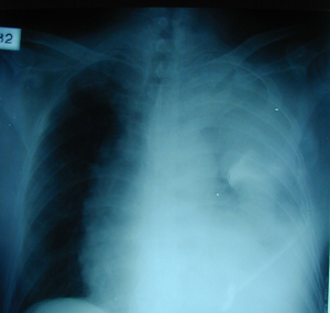
Chest X-ray after an unsuccessful effort to evacuate the fluid, which was confirmed to be fresh blood, by the insertion of chest tubes at different sites after the medical management of the abnormal results of the coagulation blood tests. The patient was on anti-coagulant medication as a treatment to pulmonary embolism.
Case 1
Figure 3
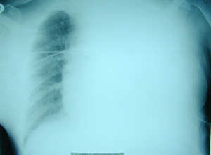
The same picture after multiple unsuccessful efforts to evacuate the blood.
Case 1
Figure 4
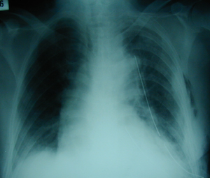
Chest X-ray after a limited left thoracotomy. Complete expansion of the lung after the removal of the blood clotts was achieved. The mediastinum is in its normal position.
Case 2
Figure 1
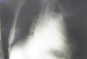
Plain postero-anterior chest X-ray after chest trauma (Fall from a large height). The left hemithorax is filled with fluid. The patient had a severe chest wall deformity.
Case 2
Figure 2
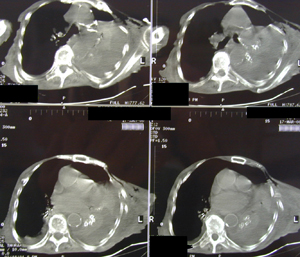
Chest CT scan. There is a large amount of blood in the left hemithorax with multiple rib fractures.
Case 2
Figure 3
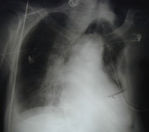
Postero-anterior chest X-ray after the insertion of a chest tube and the evacuation of a large amount of blood. There is a complete expansion of the lung, while the chest tube is visible.
Case 2
Figure 4
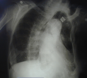
Postero-anterior chest X-ray after the evacuation of the blood, the complete expansion of the lung and removal of the chest tube.
Top