Case 1
Figure 1
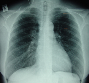
Postero-anterior chest X-ray which shows a mass at the right upper lobe of the lung, above the hilum.
Case 1
Figure 2
Lateral chest X-ray. There is a round mass at the right upper lobe above the hilum.

Case 1
Figure 3
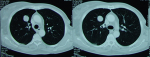
Chest CT Scan that shows a tumour mass located at the right upper lobe, which has smooth borders and without any calcification.
Case 1
Figure 4
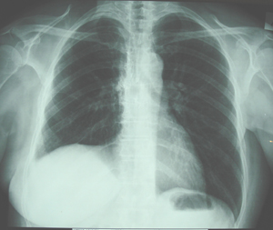
A postoperative postero-anterior chest X-ray after a right upper lobectomy. Complete expansion of the remaining middle and lower lobe.
Case 1
Figure 5
Lateral chest X-ray, after a right upper lobectomy. The histology examination of the surgical specimen showed a typical carcinoid of the lung.

Case 1
Figure 6
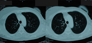
Postoperative chest CT with normal findings after right upper lobectomy.
Top