Figure 1
Postero-anterior chest X-ray, of a female patient who had a very difficult tracheal intubation for cholocystectomy operation. There is a massive pleural effusion, an air fluid level, throughout the right pleural cavity and the mediastinum.
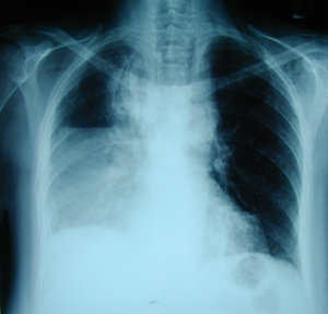
Figure 2a-d
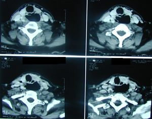
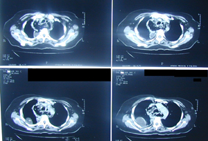
CT scan of the chest and the cervix. The results confirm the chest X-ray findings. Air fluid levels are found at the higher mediastinum, and the cervix. These radiological findings start from the cervix, running down through the mediastinum, resulting in the formation of empyema at the right hemithorax. Paracentisis of the pleural cavity revealed the existence of pus in the thoracic cavity.
A-B
C-D
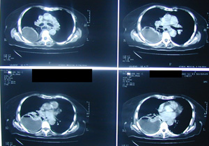
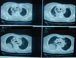
Figure 3
Postoperative chest X-ray after the performance of right thoracotomy and
wide surgical drainage of the mediastinum and the pleural cavity. Also
through a separate cervical incision the oesophageal ruptured wall was
sutured. The suture line was reinforced with the use of the sterno-clido
mastoid muscle.
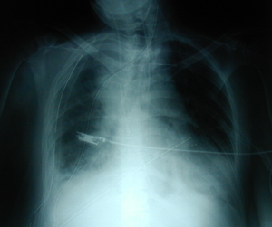
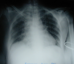
Figure 4
Postoperative chest X-ray before the patient’s discharge from the hospital after a hospitalization period of 45 days. There is complete expansion of the right lung, without any abnormality.
Top